

Diagnostic Tests
Pelvic Floor Studies
A full history and physical examination in the office by a board certified colon and rectal surgeon is the first significant step towards determining the cause of obstructed defecation and/or fecal incontinence. Based on the office visit, further studies including anal ultrasound, anal manometry, pudendal nerve terminal motor latency, defecography, dynamic MRI and/ or colonoscopy may be ordered. These studies will help your physician determine what the causes of obstructed defecation and fecal incontinence and therefore what the optimal treatment plan may be. It is not unusual for there to be more than one contributing cause to pelvic floor dysfunction. This is also true for fecal incontinence.
Anal Ultrasound
Anal Ultrasound is a study of the anal sphincter anatomy. It is performed with ultrasound waves and is painless. The test will be performed on an outpatient basis in one of the clinics at Anne Arundel Medical Center. The test will be administered by a colon & rectal surgeon. An ultrasound probe will be placed into the anal canal and ultrasound waves will be used to determine the anal sphincter anatomy- specifically if the muscles of the anal sphincter muscle complex have been damaged. Another reason for an Anal Ultrasound is to evaluate a complex perirectal fistula.
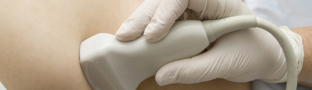
Anal Manometry
Anal Manometry is a study of the pressures generated in the anal canal. It is also a painless examination and performed on an outpatient basis in one of the Anne Arundel Medical Center clinics. A specialized catheter will be placed into the anal canal and pressure readings from the anal canal will be obtained while the anus is at rest, squeezing, straining, and coughing. The data will be used to create a 3-D map of the pressures generated in the anal canal.
Defecography
Defecography is a real-time radiologic study of defecation or the passage of a bowel movement. It is performed in the radiology department at Anne Arundel Medical Center. As part of the test, you will be asked to drink some oral contrast so the small bowel can be seen on the X-ray. You will also have a special paste placed into the rectum to simulate a bowel movement. The paste is also designed to be seen on X-ray. This study will give anatomic information about possible blockage that can occur during a bowel movement and lead to incomplete passage of stool or even trapping of stool which is expelled at a later point in time leading to passive fecal soiling.
Dynamic Pelvic Floor MRI
Dynamic MRI is also a real-time radiologic study of defecation or the passage of a bowel movement. Unlike defecography, it does not involve placement of any paste into the rectum. The study will give anatomic information about what occurs structurally during a bowel movement. It can also evaluate additional pelvic structures such as the bladder and the uterus that can also contribute to pelvic floor dysfunction.
Colonoscopy
Colonoscopy is an endoscopic evaluation of your colon. It involves a bowel preparation to be completed the day before the colonoscopy. The colonoscopy is generally performed under sedation- what many term “twilight anesthesia”. This is an important test to ensure that a new complaint of fecal incontinence is not caused by a cancer. If you have already had a recent colonoscopy, it may not be necessary.
Staging for Colon and Rectal Cancer
Once a diagnosis of Colorectal cancer has been made, it is important to stage the cancer. This is especially vital in the treatment of rectal cancers. All colorectal cancers are staged on the TNM system. T stands for tumor- the first location (primary) where the cancer occurs and source of any spread to other locations. N stands for lymph Nodes. Colon and rectal cancers are adenocarcinomas and therefore have a typical pattern of discontinuous spread to lymph nodes first. M stands for metastasis. Technically, the first spread to lymph nodes is also considered metastasis. Since the local lymph nodes are removed at the time of surgery, they are regarded differently than spread to other organs such as the liver, the lungs or the brain. Staging of cancer at the time of diagnosis helps the physicians determine treatment strategy. In the case of rectal cancer, a rectal ultrasound or a pelvic MRI will provide the T and N stages. MRI or CT scan, usually in combination with a PET scan will be used to provide limited information about the primary tumor, and lymph nodes. Their main role is to identify metastatic disease.
Rectal Ultrasound
A rectal ultrasound is a study looking at the wall of the rectum and surrounding lymph nodes using high-frequency sound waves, or ultrasound usually for the purpose of initial rectal cancer staging, follow up for a history of rectal cancer, or evaluation of a rectal carcinoid. The test takes about 20 minutes and is performed on an outpatient basis in one of the clinics at Anne Arundel Medical Center. This study is performed in conjunction with a rigid proctoscopy. Once the proctoscope has been inserted to the level of the upper rectum, the ultrasound probe is placed through the proctoscope and the ultrasound waves will be used to evaluate the rectal anatomy.
In the case of rectal cancer staging, the focus of the examination will be determining how deep into the rectal wall the primary cancer has invaded, and if the cancer has penetrated neighboring organs, such as the prostate gland in men or the vagina in women. This will determine the T, or tumor stage. The local lymph nodes will also be evaluated for evidence of spread, or metastasis, which will determine the N, or lymph node stage. Rectal ultrasound is not able to assess distant spread to organs such as the liver, lungs or brain since it is only looking at the rectum and surrounding tissues.
CT Scan of the abdomen and pelvis
A CT (computed tomography) scan or CAT (computed axial tomography) scan is an x-ray study of the body, typically the abdomen and pelvis to evaluate a variety of complaints and disease states. Unlike traditional x-rays, a CT will provide detailed anatomic information about all of the abdominal and pelvic organs. It is a standard part of pre-treatment staging for colorectal cancer among other diseases and is typically combined with a PET scan. The test is frequently administered with an iodine-based, IV contrast as well as an oral contrast. If you have a known allergy to iodine or shellfish, have impaired kidney function or take oral hypoglycemic, such as metformin for diabetes it is important to inform your doctor prior to having the test scheduled. A CT scan is frequently performed on an outpatient basis, although hospital in-patients can also have it done.
PET Scan
A positron emission tomography is a study generally used to assist in staging of colorectal cancers. A radioactively labeled sugar molecule called FDG is used as a tracer molecule to detect cells with an increased level of metabolic activity. These cells will take up a disproportionate amount of the tracer causing them to “light-up” on the 3-D image of the body. This technique is often combined with a CT scan image done on the same day to improve diagnostic accuracy.
Pelvic MRI
MRI stands for magnetic resonance imaging. Like with ultrasounds, there is no radiation given with this test. A powerful magnet is used to align the atomic nuclei in the body, typically focusing on water and fat content in the various tissues. Like a CT scan, the MRI is typically used to provide detailed images of the abdominal and pelvic organs and structures. It can be of particular use in staging of rectal cancers and in some institutions has replaced the rectal ultrasound for staging. In rare cases it is also used to look for recurrence of rectal cancers after initial treatment. If you have claustrophobia, you need to discuss this with the doctor before your test is scheduled.
 For more information on our Diagnostic Tests, or to schedule an appointment, please complete our online form or call 410-573-1699.